Medical Imaging Breakthrough: How Xenon Gas Enhances MRI and CT Scan Precision for Pulmonary Diagnostics
BY Tao, Published Feb 1, 2026
In the world of medical diagnostics, the human lung has long remained a “dark continent.” While we have mastered the art of imaging bones, brains, and solid organs with exquisite detail, the lung—a sponge-like organ filled mostly with air—has historically frustrated our best technologies. Standard X-rays offer only 2D shadows; CT scans provide excellent structural maps but tell us little about function; and conventional MRI struggles to image air-filled voids.
However, a revolution is quietly taking place in radiology departments and research centers worldwide. It is driven not by a new machine, but by a new material: Xenon (Xe).
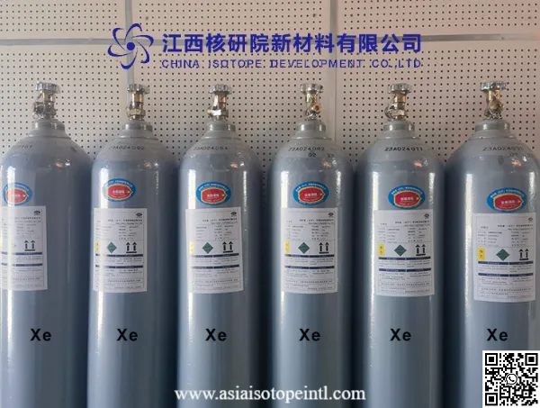
As China Isotope Development Co Ltd was supplying High Purity Xenon Gas to world’ clients, I have watched Xenon evolve from a niche industrial component into one of the most exciting contrast agents in modern medicine. Specifically, Hyperpolarized Xenon-129 (Hp-Xe) for MRI and Xenon-Enhanced CT are fundamentally changing how we diagnose pulmonary diseases—from COPD and asthma to the mysterious lingering effects of Long COVID.
In this article, I will take you inside the physics, physiology, and clinical application of this breakthrough, explaining why this heavy, inert gas is becoming the gold standard for precision pulmonary diagnostics.
1. The “Black Hole” of Lung Imaging: Why We Need Xenon
To understand the value of Xenon, we must first understand the limitations of current technology.
The MRI Problem: Magnetic Resonance Imaging (MRI) works by detecting the protons (hydrogen nuclei) in water molecules within our bodies. Since the human body is mostly water, MRI works beautifully for the brain, liver, and muscles. The lungs, however, are different. They are essentially bags of air with very low tissue density. There are simply not enough protons in the lungs to generate a strong signal. On a standard MRI, healthy lungs appear as a black void—a signal “black hole.”
The CT Problem: Computed Tomography (CT) uses X-rays to create detailed 3D pictures. While CT is excellent at showing structure (scarring, tumors, collapsed airways), it is a static image. It cannot easily show function. It cannot tell you if oxygen is actually crossing from the air sac into the bloodstream. A patient can have a “normal” looking CT scan but still suffer from severe breathlessness.
The Spirometry Problem: The standard breathing test (blowing into a tube) gives us global numbers, like FEV1 (Forced Expiratory Volume). It tells us that the lung is failing, but not where or why. It is an average measurement that masks regional defects.
The Xenon Solution: By introducing Xenon gas as an inhaled contrast agent, we illuminate the darkness. We turn the lungs from a black void into a bright, detailed map of ventilation and gas exchange.
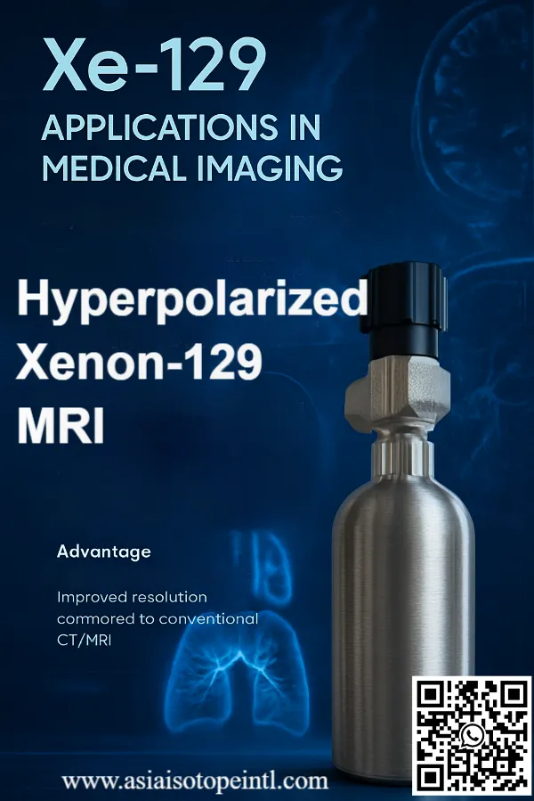
2. Hyperpolarized Xenon-129 MRI: The Quantum Leap
The most cutting-edge application involves a specific isotope: Xenon-129. But we don’t just use standard Xenon-129; we “hyperpolarize” it. This is where high-level physics meets clinical medicine.
The Physics of Spin Exchange Optical Pumping (SEOP)
In a standard MRI, a powerful magnet aligns the “spins” of protons in your body. Only a tiny fraction of them align, creating a relatively weak signal that requires the machine to listen carefully.
To make Xenon visible, we use a process called Spin Exchange Optical Pumping (SEOP).
- We take a mixture of Xenon-129, Nitrogen, and Helium.
- We introduce alkali metal vapor (usually Rubidium).
- We shine a circularly polarized laser light into the mixture. The laser aligns the electrons of the Rubidium.
- Through collisions, the Rubidium transfers this alignment (polarization) to the nucleus of the Xenon-129 atoms.
The result? The Xenon gas becomes hyperpolarized. Its magnetic signal is amplified by a factor of 100,000 times compared to thermal equilibrium.
When a patient inhales a single breath of this hyperpolarized gas and holds it for 10-15 seconds, the MRI scanner picks up a blazing bright signal. We are no longer looking for water protons; we are imaging the gas itself.
3. The Three Maps: Seeing Physiology in Real-Time
What makes Xenon-129 MRI truly revolutionary is that it doesn’t just show us where the air goes. Because Xenon is soluble in lipids and tissue, it allows us to track the entire journey of a breath.
We can generate three distinct “maps” from a single breath-hold scan:
A. The Ventilation Map (Where the air is)
This image shows exactly which parts of the lung are receiving air and which are not. In conditions like asthma or emphysema, we see “ventilation defects”—dark patches where air is trapped or blocked. Unlike spirometry, which gives a global number, this map shows the specific location of the problem.
B. The Barrier Uptake Map (Crossing the membrane)
This is the game-changer. Xenon dissolves into the delicate membrane (the interstitial tissue) separating the air sac (alveolus) from the blood. Xenon-129 has a unique chemical property: its magnetic frequency shifts slightly (called a chemical shift) when it moves from gas to dissolved tissue. The MRI scanner can “tune” into this specific frequency. We can literally generate an image showing the health of the lung tissue itself. If the membrane is thickened by fibrosis (scarring), the signal changes.
C. The Red Blood Cell (RBC) Map (Entering the stream)
Finally, Xenon diffuses from the membrane into the red blood cells, binding momentarily to hemoglobin. Again, the magnetic frequency shifts. We can image this specific frequency to see if oxygen (proxied by Xenon) is successfully reaching the blood. This is critical for diagnosing diseases like Pulmonary Hypertension, where the airways are open (good ventilation) but the blood vessels are blocked or damaged (poor RBC transfer).
4. Clinical Applications: Solving Medical Mysteries
As a researcher, I am most excited about how this technology is solving diagnostic dilemmas that have baffled doctors for years.
Long COVID and Unexplained Dyspnea
Since 2020, millions have suffered from “Long COVID,” complaining of severe shortness of breath despite having normal CT scans and normal spirometry. This has been frustrating for both patients and doctors. Recent studies using Xenon-129 MRI have revealed the “invisible” cause. These scans often show RBC transfer defects. The air is getting in, but oxygen isn’t getting into the blood efficiently, likely due to microscopic clots or subtle inflammation in the capillaries that standard CT scans simply cannot see. Xenon validated the patients’ symptoms when other tests failed.
COPD and Emphysema
In Chronic Obstructive Pulmonary Disease (COPD), Xenon MRI is more sensitive than CT. It can detect early-stage emphysema and small airway disease before significant structural damage occurs. This allows for earlier intervention and better monitoring of drug therapies.
Asthma
Asthma is a heterogeneous disease; it moves around the lungs. Xenon MRI allows researchers to see “ventilation heterogeneity”—patchy areas of airflow obstruction. It is being used to test new bronchodilator drugs to see exactly how well they open up specific regions of the lung.
Idiopathic Pulmonary Fibrosis (IPF)
In fibrosis, the lung tissue thickens and scars. The “Barrier Uptake Map” generated by Xenon MRI provides a direct measurement of this thickening. It offers a non-invasive way to track whether anti-fibrotic drugs are working, sparing patients from risky lung biopsies.
5. Xenon-Enhanced CT (Xe-CT): The Structural Contrast Agent
While Hyperpolarized MRI gets the headlines for its functional mapping, Xenon-Enhanced Computed Tomography (Xe-CT) plays a different but equally vital role.
The Mechanism: Xenon as a Radiopaque Gas
In CT scanning, we typically use iodine-based liquids injected into the vein to create contrast. However, you cannot inject liquid iodine into the airways. Xenon has a high atomic number (54), which means it is dense with electrons. These electrons absorb X-rays efficiently, making Xenon radiopaque. When a patient inhales a mixture of Xenon (typically 30%) and Oxygen, the gas appears bright white on a CT scan.
Applications of Xe-CT
- Regional Ventilation Measurement: Similar to MRI, Xe-CT can show ventilation defects. It is higher resolution than the traditional nuclear medicine “V/Q scan” (Ventilation/Perfusion) and does not require radioactive isotopes like Technetium-99m.
- Cerebral Blood Flow (CBF): This is a unique application. Because Xenon is lipid-soluble, it crosses the blood-brain barrier freely. By inhaling Xenon while undergoing a CT scan of the head, doctors can measure blood flow to different parts of the brain quantitatively. This has been used in stroke assessment and traumatic brain injury management.
6. Safety and Physiology: Why Xenon is Ideal
One might ask: “Is it safe to inhale a heavy noble gas?” The answer is an emphatic yes, provided it is mixed with oxygen. In fact, Xenon has been used as a general anesthetic for over 60 years.
- Non-Toxic: It is chemically inert. It does not react with the body’s tissues to form toxins.
- Rapid Elimination: Because it is a gas with low blood solubility, it is exhaled rapidly once the patient stops inhaling it. Most Xenon leaves the body within minutes.
- Neuroprotection: Interestingly, Xenon has neuroprotective properties. It inhibits NMDA receptors in the brain, which can protect neurons from injury during strokes or trauma.
- No Radiation (MRI): Unlike CT or V/Q scans, Xenon MRI involves zero ionizing radiation. This makes it safe for repeated use in children (e.g., for Cystic Fibrosis monitoring) and for longitudinal studies where patients need to be scanned many times over years.
7. The Technical and Supply Chain Challenges
As an expert in specialty gases, I must address the logistical reality. Implementing Xenon imaging is not as simple as buying a new software update. It involves sophisticated gas engineering.
Isotope Enrichment
For MRI, we need Xenon-129. Natural Xenon contains only about 26% Xe-129. While natural Xenon can be used, many advanced research applications prefer enriched Xenon (80-90% Xe-129) to boost the signal even further. Producing this requires gas centrifuges or thermal diffusion columns—complex separation technologies.
The Polarizer
Hospitals need an on-site “polarizer”—the machine that uses lasers and Rubidium to hyperpolarize the gas. These are complex devices, though recent years have seen the development of compact, FDA-approved clinical polarizers that fit easily in a hospital radiology suite.
Cost and Recovery
Xenon is expensive. It is a rare byproduct of air separation (fractional distillation of liquid oxygen). A single lung scan might use a few liters of gas. To make this economically viable, we use Xenon Recovery Systems. These units capture the patient’s exhaled breath, remove moisture and CO2, and cryogenically trap the Xenon so it can be purified and reused (for non-medical or re-sterilized applications).
8. The Future: Where Do We Go From Here?
The potential of Xenon imaging extends far beyond the lungs. Research is currently exploding in several new directions:
1. Brown Fat and Metabolism: Brown Adipose Tissue (BAT) helps regulate body temperature and metabolism. Xenon dissolves readily in fat. Researchers are using Xenon MRI to image brown fat activity, which could offer insights into obesity and metabolic disorders.
2. Brain Imaging: Because dissolved Xenon-129 has a specific spectral signature in brain tissue, researchers are developing techniques to image “brain uptake” of Xenon. This could lead to new ways to diagnose Alzheimer’s, vascular dementia, and traumatic brain injury by revealing subtle changes in brain tissue composition and perfusion.
3. Kidney Imaging: Early work suggests Xenon can image renal blood flow and tissue oxygenation, offering a non-invasive way to assess kidney failure risks.
4. Pharmaceutical Trials: Pharmaceutical companies are increasingly using Xenon MRI as a biomarker in clinical trials. Instead of waiting years to see if a lung drug improves survival rates, they can see in weeks if the “Barrier Uptake” signal is improving. This accelerates drug development and lowers costs.
Conclusion: Lighting the Way
We are witnessing a paradigm shift in pulmonology and radiology. For decades, doctors have treated lung diseases by listening to sounds through a stethoscope and looking at shadows on an X-ray.
Xenon gas changes everything. It transforms the lung from a static black-and-white image into a vibrant, technicolor map of physiology. It allows us to see the air entering the bronchi, crossing the alveoli, and dissolving into the blood. It allows us to detect disease before it destroys tissue.
From the physics of spin exchange optical pumping to the bedside diagnosis of a post-COVID patient, Xenon represents the perfect marriage of basic science and clinical care. As a specialty gas expert, I am proud to see this noble element—once considered merely “inert”—playing such an active and vital role in saving lives.
The future of medical imaging is not just digital; it is gaseous. And it is being illuminated by Xenon.
Would you like a deeper dive into any specific technical parameters or applications?
As an industry leader focused in Specialty Gases products, our goal is to support our customers by keeping them at the forefront of their industries. We’re here to help with any questions you might have so you can transform your ideas into reality, and tackle those big science challenges.
Get free consultant, our experts are ready to serve.
(Follow up our update articles on www.asiaisotopeintl.com or send your comments to tao.hu@asiaisotope.com for further communications)